
To get started with Image Review, choose from the list below:

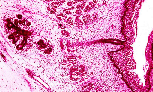
The bones of the vault of the skull and of the face (including most of the mandible) develop by the process of intramembranous bone formation in the embryo. In this low power photomicrograph, a portion of the developing mandible is illustrated. A branched spicule of bone can be seen near the center of the field. Osteoblasts line the surface of the developing bone, and it is surrounded by loose mesenchyme. A more condensed layer of mesenchyme, the developing periosteum, surrounds the bone. Developing muscle can be seen at the right, apparently attaching to the periosteum. Within the spicule of bone, the homogeneous pink material is osteoid (prebone) while the darker purple material is calcified (mineralized) bone matrix.
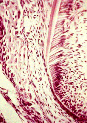
Cementum
The Cementum is the part of the periodontium that attaches the teeth to the alveolar bone by anchoring the periodontal ligament.
Dentin & Pulp
Dentin is a calcified tissue of the body, and along with enamel, cementum, and pulp is one of the four major components of teeth.
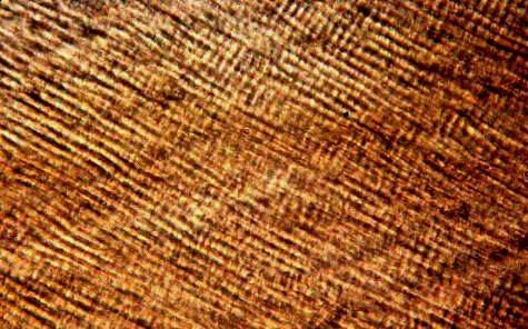
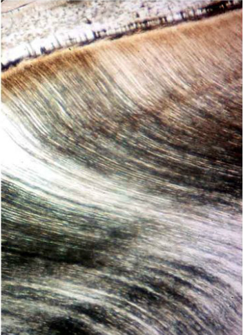
Structure of Enamel
Enamel is the hardest substance in the human body and contains the highest percentage of minerals.
Facial Growth & TMJ
Facial Growth issues can create undue stress over time to the Tempromandibular joint (TMJ) causing inflammation.
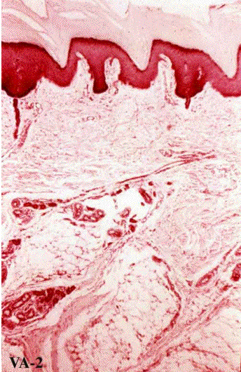
Oral Mucous Membrane
The oral mucous is the membrane lining the inside of the mouth.
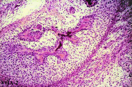
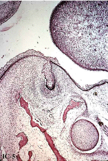
Gingiva & Dentogingival Junction
Gingiva are part of the soft tissue lining of th mouth. The Dentogingival Juntion holds in place the junctional or attachment epithelium.
Enamel Formation
Amelogenesis is the formation of enamel on teeth and begins when the crown is forming during the bell stage of tooth development.
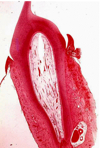
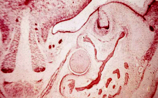
Development of Teeth
Referred to as odontogenesis is the complex process by which teeth frorm from embryonic cells, grow, and erupt into the mouth..
Salivary Glands
Are exocrine glands, glands with ducts, that produce saliva. The also secrete amylase, an enzyme that breaks down starch into maltose.
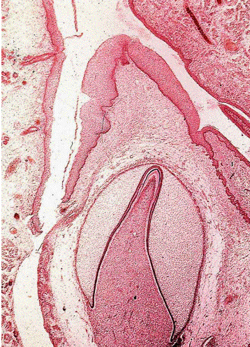
Eruption & The Alveolar Process
Tooth Eruption is a process in tooth development in which the teeth enter the mouth and become visible.
